Our Services

Services We Provide

EMG Electromyography Testing

Electromyography (EMG) is a diagnostic procedure that evaluates the health of muscles and the nerve cells that control them. This test measures the electrical activity produced by the muscles during contraction and relaxation. It helps in identifying conditions such as muscle disorders, nerve disorders, and disorders affecting the nerve roots.
During an EMG, a small needle electrode is inserted into the muscle to be tested, and the electrical activity is recorded and analyzed. The procedure is performed by a trained healthcare professional, usually a neurologist or a physiatrist. It is safe and minimally invasive, and the results provide valuable information for diagnosing and managing a variety of conditions affecting the muscles and nerves.
A nerve conduction study (NCS) is often performed in conjunction with an electromyography (EMG) to assess the health and function of nerves. During an NCS, small electrical pulses are applied to the nerves, and the response is recorded. This helps in determining the speed and strength of nerve signals, as well as identifying potential nerve damage or dysfunction. NCS can help diagnose conditions such as carpal tunnel syndrome, peripheral neuropathy, and nerve root compression. It is a safe and minimally invasive procedure that provides valuable information about nerve health and function.

Preparing for an EMG:

  • Speak with your neurologist about any medications you are taking, as some medications may need to be adjusted or stopped before the procedure.
  • Avoid using lotions or oils on your skin the day of the test, as they can interfere with the placement of the electrodes.
  • Tell your neurologist if you have a pacemaker or other implantable device, as this may affect the procedure.
  • Wear comfortable, loose-fitting clothing that allows access to the muscles being tested.

Before the procedure:

  • Your neurologist will review your medical history and ask about any symptoms you may be experiencing.
  • You may be asked to change into a hospital gown or other clothing that allows access to the muscles being tested.
  • You will be positioned comfortably on an exam table or bed.
  • The skin over the muscles being tested will be cleaned with an antiseptic solution.
  • A small, thin needle electrode will be inserted into the muscle being tested, and you will be asked to contract and relax the muscle several times while the electrical activity is recorded.
  • The needle electrode will be removed, and the procedure will be complete.

EEG Electroencephalogram Testing

An Electroencephalogram (EEG) is a non-invasive test that measures and records the electrical activity of the brain. This test involves placing small electrodes on the scalp, which are connected to an EEG machine that amplifies and displays the brain’s electrical signals as wave patterns. An EEG helps in diagnosing various neurological disorders, such as epilepsy, sleep disorders, and brain tumors. It is also used to monitor brain activity during surgeries and to assess brain function in cases of brain injury or coma. EEG is painless and safe, and the results provide valuable information for diagnosing and managing neurological conditions.

Preparing for an EMG:

  • Speak with your neurologist about any medications you are taking, as some medications may need to be adjusted or stopped before the procedure.
  • Avoid consuming caffeine and alcoholic beverages for at least 24 hours before the test.
  • Avoid using hair products such as gels, hairsprays, and oils, as they can interfere with the placement of the electrodes.
  • Wash your hair the night before the test, and avoid using conditioner or other hair products that can leave residue on your scalp.
  • Get plenty of rest the night before the test, as sleep deprivation can affect the results.

Before the procedure:

  • You will be asked to remove any metal objects from your body, including jewelry, eyeglasses, and hairpins.
  • You may be asked to wear a hospital gown or other clothing that allows easy access to your head and neck.
  • The technician will measure the circumference of your head and mark the locations where the electrodes will be placed.
  • The technician will use a special paste or glue to attach the electrodes to your scalp, and the EEG recording will begin. The test typically takes about 30-60 minutes, during which you will be asked to relax and close your eyes for some portions of the test.

After the procedure:

  • The electrodes will be removed, and the paste or glue will be washed off your scalp.
  • You can resume your normal activities immediately after the test.
  • Your neurologist will review the results with you and discuss any further testing or treatment that may be needed.

Botox Injection

Botox, or botulinum toxin, is a medication used for a variety of neurological conditions. It is most commonly known for its use in cosmetic procedures to reduce the appearance of wrinkles and fine lines. However, Botox can also be an effective treatment for certain neurological disorders, including:

Chronic Migraines: Botox injections can be used as a preventive treatment for people who experience chronic migraines, defined as headaches that occur on 15 or more days per month. Botox can help reduce the frequency and severity of migraines by blocking the release of certain chemicals involved in pain transmission.
Cervical Dystonia: Also known as spasmodic torticollis, cervical dystonia is a condition characterized by involuntary contractions of the neck muscles, causing the head to twist or turn in an abnormal position. Botox injections can help relax the affected muscles, reducing pain and improving range of motion.
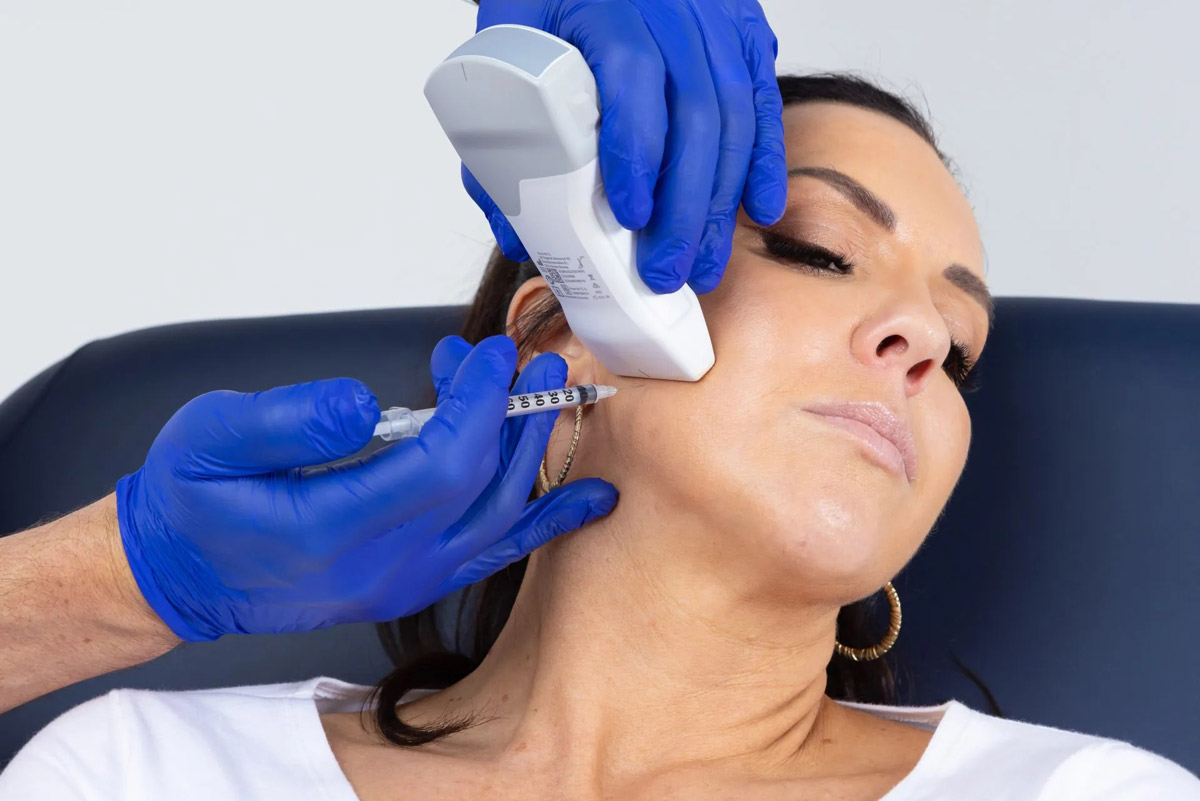
Hemifacial Spasm: This condition involves involuntary contractions or twitching of the facial muscles on one side of the face. Botox injections can help reduce the frequency and severity of these spasms, improving quality of life for individuals with hemifacial spasm.

Blepharospasm: Blepharospasm is a condition characterized by involuntary contractions of the muscles around the eyes, causing excessive blinking or difficulty opening the eyes. Botox injections can help relax these muscles, reducing blinking and improving vision.
It’s important to note that Botox injections are a temporary treatment, and the effects typically last for a few months. Repeat injections may be needed to maintain the desired results. Additionally, Botox injections may not be suitable for everyone, and potential risks and side effects should be discussed with your neurologist before undergoing treatment.